Cone Beam Computed Tomography

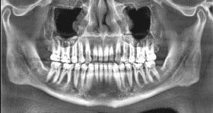
All navigation starts with a map. Cone Beam 3-D images provide high-definition, three-dimensional, digital data, and precise anatomical information of all oral and maxillofacial structures. These 3-D images are the map that our doctors use to plan and execute your surgical treatment. They are the key to all image guided surgery. This is done: With less radiation then conventional Medical CT. In a comfortable “open”......
Read more »

Static Image Guided Surgery

Static Guided Surgery is done primarily to place dental implants. Static Image Guided Implant Surgery involves a number of steps. A CBCT is taken of your jaw to evaluate proposed implant site. An intraoral laser scan (iTero) is taken of your jaws. This new technology allows a virtual model of your jaw to be superimposed on your 3-D CBCT for planning purposes. Your dentist and......
Read more »

X-Guide Navigated Surgery – XNAV

Dynamic Image Guided Surgery (Stereotactic Surgery) is a new and exciting field of dental surgery. Dr. Emery is one of the inventors of the X-Guide. This type of surgery was pioneered in medicine. It is used by neurosurgeons and other medical surgical specialties to allow the surgeon to “virtually” see into their patient real time while they operate and “guide” their instruments to accurate locations......
Read more »

What is Image Guided Surgery?

In a traditional dental implant placement, a doctor must mentally visualize aspects of the surgery based on diagnostic tests and then conduct the procedure freehand. This increases the risk that the surgery will deviate from the ideal placement, and patients might not get the most aesthetic and durable outcome. As an alternative, Capital Center® for Oral and Maxillofacial Surgery offers guided implant surgery to patients.......
Read more »