Technology in the field of implant dentistry has come a long way in the past decade, with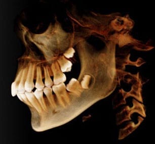 advancements like 3D imaging and image guided surgery making implant placement planning and procedures safer and more accurate. By putting these cutting-edge technologies to use in the placement of dental implants, oral surgeons Dr. Robert Emery is also able to offer patients quicker and more comfortable procedures than was possible years ago. Read on to learn what these technologies are and how they have streamlined and improved the dental implant process.
Dental Implants and i-CAT 3-D Imaging
3-D imaging is one of the most important advances in dental technology, offering dental professionals the ability to examine oral structure and anatomy in much better detail than traditional X-ray imaging can. Capital Center for Oral and Maxillofacial Surgery uses i-CAT cone beam technology to produce high-definition, three dimensional digital images of all oral and maxillofacial structures and anatomy, making possible more precise evaluations of factors crucial to the most accurate placement of dental implants.
We can, with these images, evaluate bone quantity and quality, determine the exact location of nerves and sinus cavities, and make accurate, distortion free measurements for greater surgical predictability.
Another benefit of 3-D imaging that patients are sure to appreciate is the comfort and speed of the imaging process as compared to traditional X-rays. Scanning with i-CAT takes less than ten seconds to complete in most cases, is done in a comfortable, open environment and involves very low radiation levels, with exposure ten times less than a typical hospital CT scan.
Dental Implants: Image Guided Dental Implant Procedures
Specialized software enables the digital images created by i-CAT 3D imaging to be transformed into an interactive digital model of a patient’s oral and maxillofacial anatomy, enabling the creation of precise, dynamic surgical guides.
In image guided surgery, those surgical guides are implemented in real time, using on-the-spot images of bone and tissue to provide surgeons with a virtual navigator to indicate the optimal position for the placement of each dental implant. The exceptional level of precision offered by image guided surgery allows implant placement with smaller incisions, shorter procedure duration, less patient discomfort and decreased recovery time.
Capital Center for Oral and Maxillofacial Surgery is the first and only surgical center in the D.C area to offer a full range of image 3-D image guided surgeries, including image guided placement of dental implants. Patients will find that these technologies make dental implant restorations easier, safer and more accurate than ever before. So if your smile is in need of a little work, there has never been a better time to have that restoration done.
Request a consultation to see how our board certified oral and maxillofacial surgeons can use today’s exceptional technology to create your perfect smile.